Cone Beam Computed Tomography
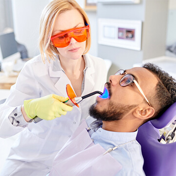
When we go to the dentist, we all want to know that we are getting the best treatment possible. While our dentists always do their best to find any issues lurking inside our mouths, they sometimes need help to spot particular issues. Most of us know the importance of x-rays for diagnosing and treating different problems, but sometimes a plain old x-ray isn’t enough to do the job.
Thankfully, 3D imaging in dentistry has come a long way, and dentists have the tools to find even the most difficult-to-spot problems that a visual exam and traditional x-ray would not find. One tool at your dentist’s disposal is Cone Beam Computed Tomography or CBCT.
How Does CBCT Work?
Cone beam computed tomography may be a mouthful, but it’s an advanced form of x-ray technology that creates a 3D image of the inside of a patient’s mouth all at once. This is much more effective and easier than taking several flat x-rays. CBCT uses a cone-shaped x-ray beam that rotates on a fixed point to create one solid image. The image is then fed to a computer, which can be manipulated and enhanced using the accompanying software.
This means that dentists can get the full picture of a patient’s mouth in one image, including areas that would normally be hard to see. That image can be enhanced and modified by adding computer technology to look for certain problems. Your dentist can focus on the teeth, the gums, or the soft tissues inside the mouth when trying to determine if something is wrong such as gum disease, tooth decay, or even signs of cancerous tissue.
This means faster work for the dentist, less time in the dental chair for patients, and a more positive outcome when finding and treating a wide range of oral health issues during a routine dental clinic visit.
Author's Bio:

Dr. Darryl Azouz is committed to providing gentle, holistic pediatric dental care that supports both oral health and overall wellness. He focuses on non-toxic treatments, early prevention, and creating a positive dental experience for children. His approach ensures healthy development while minimizing exposure to harmful materials. He also educates parents on nutrition and lifestyle habits that influence oral health. By fostering trust and comfort, he helps children build lifelong healthy dental habits.